
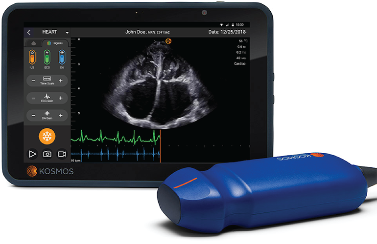
EchoNous, a developer of novel ultrasounds, has found a way to leverage multiple critical clinical technologies within a single device. The result is KOSMOS, a handheld 3-in-1 device consisting of an ultrasound, electronic stethoscope, and an ECG, all linked via artificial technology.
When COVID-19 was hitting NYC earlier this year, EchoNous was able to use KOSMOS to help diagnose COVID patients and to inform on treatment strategies. We had a lovely chat with Dr. Richard Hoppmann, a key member of EchoNous, Clinical Professor of Internal Medicine at the University of South Carolina, and the Director of the Ultrasound Institute.
Alice Ferng, Medgadget: Can you tell me a bit about yourself and how you got interested in ultrasound technologies that led to your involvement with EchoNous?

Dr. Richard Hoppmann, innovator at EchoNous: I am an internist/rheumatologist by training and have been with University of South Carolina School of Medicine since 1990. My career here has included Directorship of the Division of Rheumatology, Chief of Medical Service at the Dorn VA Hospital, Chair of the Department of Internal Medicine, Associate Dean of Medical Education, Dean of the School of Medicine, and Director of the Ultrasound Institute.
In 2006, while Associate Dean of Medical Education, we initiated the first-in-the-nation integrated ultrasound curriculum for medical students across all four years of medical school. We subsequently founded the first professional organization dedicated to ultrasound in medical education – the Society of Ultrasound in Medical Education (SUSME) – and hosted the first World Congress on Ultrasound in Medical Education in 2011. There have been six other world congresses since then with the most recent being at the University of California-Irvine. The 2020 Congress had to be rescheduled due to COVID and is now scheduled at Wayne State University School of Medicine in Detroit in August of 2021. We have expanded our ultrasound program over the past 15 years to include training residents, physician assistants, practicing physicians, and others. Additional information on our programs can be provided.
Our focus has been primarily on developing ultrasound as a learning tool and as a frontline healthcare provider clinical practice tool. We have done this by developing educational material for our medical students as well as providing it to others via open access on our Ultrasound Institute website. Our Youtube video channel on ultrasound scanning has had over 1.3 million views worldwide. We have hosted many conferences and have been working with many medical schools nationally and internationally to develop ultrasound curricula. We have also partnered with national and international organizations to advance ultrasound education such as the American Institute of Ultrasound in Medicine (AIUM) and the World Interactive Network Focused on Critical Ultrasound (WINFOCUS).
Approximately 12 years ago we began looking for innovative ways to enhance ultrasound as a teaching tool and started working with our intellectual property office on patents. We now have four patents that have been issued and have seven patents that are pending.
One of our early patents was combining stethoscope sounds (auscultation) and ultrasound in real-time. The addition of sound to ultrasound was quickly recognized as a great teaching tool, especially for understanding and assessing the heart. Our group at the University of South Carolina School of Medicine Ultrasound Institute has also been interested in artificial intelligence (AI) and how it might be used in both medical education and medical practice.
Thus, we were delighted in 2016, when Kevin Goodwin and Niko Pagoulatos from EchoNous approached us about the stethoscope sound-ultrasound patent. They had already conceived of the three-signal approach of ultrasound, auscultation, and ECG combination augmented with AI.
Medgadget: Let’s talk more about the KOSMOS device. What was the inspiration and reasoning behind incorporating ultrasound, stethoscope auscultation, and ECG in one device? What kind of information does this provide the physician?
Dr. Hoppmann: From the combination of stethoscope sounds and ultrasound perspective, I distinctly remember being in my office one evening and thinking, “I wonder what it would be like to listen to my heart with a stethoscope as I scan it with ultrasound”. So, I got my stethoscope and a portable ultrasound system and did just that. When I had the stethoscope and ultrasound probe in place over my heart and looked at the ultrasound screen I said something like “Wow, I can both see and hear my heart and how the closure of the heart valves are creating the heart sounds”.
I have always been a teacher and I knew at that moment that students would love this approach and it would give them an understanding of the heart and heart sounds that had never before been possible. So, I got to work with Shaun Riffle, an incredibly talented media expert, and we planned a session to simultaneously record the ultrasound of the heart and the stethoscope heart sounds. We recorded the heart sounds by cutting the tubing of an old stethoscope and slipping a lavalier microphone down the remaining tubing and sealed it off. When we captured our first ultrasound-heart sounds recording, we knew we had something unique. We submitted a patent on the combination and began exploring other innovative approaches to medical education and clinical practice, especially those related to ultrasound.
For us, AI is about combining the power of big data with the advances in computer processing to analyze with algorithms and neural networks huge amounts of data to significantly improve education and the practice of medicine. Kosmos is able to improve the acquisition and the accuracy of interpretation of the three signals of ultrasound, auscultation, and ECG’s individually and then in combination to provide important clinical information that is greater than the sum of the separate signals. This can help us understand disease processes and improve patient care in general as well as for the individual patient. It allows for a more personalized or precision medicine approach to the care of the patient. As an added benefit, using a single device with AI is more efficient than the traditional approach of performing and analyzing these three signals one at a time. This efficiency saves time that can then be used by the busy healthcare provider for direct personal communication with the patient which is essential in truly caring for the patient and addressing his/her healthcare concerns.
To appreciate the potential magnitude of this multi-signal AI approach, one can think of Kosmos as the smart stethoscope for the 21st century. If everyone who presently uses a stethoscope today – physicians, nurses, nurse practitioners, physician assistants, medics, and others – had access to a smart device like Kosmos with the power to capture and analyze three of the most fundamental health assessment signals in medicine with AI assistance, the world of healthcare would change dramatically. One need only reflect on how smart phones have fundamentally changed our daily lives to get a sense of the potential of Kosmos to change medical education and healthcare across the globe.
Medgadget: What kind of data are you able to obtain with KOSMOS that the traditional technologies alone did not provide?
Dr. Hoppmann: Here’s some of what Kosmos is able to do:
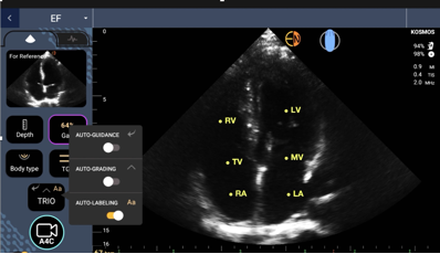
- Real-time auto-labeling of the structures being scanned such as identifying the right and left atria and ventricles of the heart. This will greatly enhance self-directed learning of ultrasound and also learning cardiac anatomy.
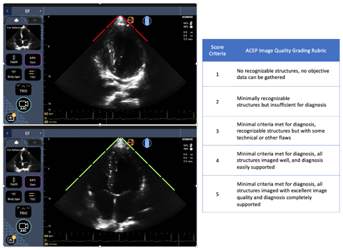
- Real-time auto-grading of the quality of image obtained on a scale from 1 to 5. This will accelerate the learner’s ability to capture quality images and also provide a standard of image quality so learners can eventually grade the quality of their own images as well as images of others. Obtaining quality images is essential to accurately determining physiological functions such as the ejection fraction of the heart.
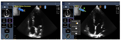
- Auto-guidance in the form of probe instructions such as “rotate the probe clockwise” are displayed in real-time on the screen thus allowing even the novice ultrasound user to quickly learn to capture quality images – another aid in self-directed learning.
- Automated calculations of important physiological measures of function such as the ejection fraction or percent of blood ejected from the heart with each beat. The ejection fraction is an excellent assessment of heart function.
- Also included is heart rate and in the future it will include ECG interpretation and heart sound interpretation.
Medgadget: How does AI play into this? Can you talk more specifically about how AI amplifies each of the three technologies, as well as how it works to bring them together for a hybrid output that provides physicians a novel way of looking at data? How are you training or developing the algorithms?
Dr. Hoppmann: The founders of EchoNous are pioneers in point-of-care ultrasound and have brought extensive expertise and success to Kosmos. In addition, they have hired experts with advanced degrees in AI, including image recognition. They also have a broad array of experienced ultrasound consultants including emergency medicine physicians, cardiologists, obstetricians, internists, and sonographers that provide additional ultrasound and clinical expertise as well as assist in the development of AI algorithms and training / testing on large populations of patients.
Training and testing of AI on large datasets enhances the diagnostic accuracy of the individual components of the three signals and the combination of data from the three signals further enhances diagnostic accuracy. This allows all users to obtain accurate images and clinical measurements that result in improved diagnosis and treatment for their patients.
Combinations of these signals allow calculation and display of important physiological measures of heart functions such as stroke volume, ejection fraction, heart rate and cardiac output.
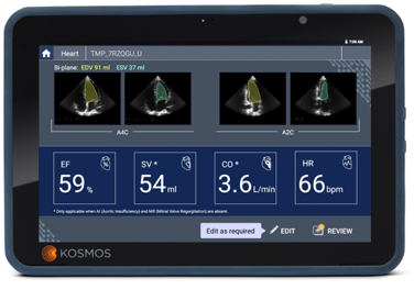
Cardiac Calculation Display: EF=ejection fraction or the percentage of blood ejected from the ventricle with each contraction, SV=stroke volume or the volume of blood ejected with each contraction or stroke of the heart, CO=cardiac output or the volume of blood pumped by the heart per minute, and HR=heart rate or beats per minute. Volume calculations are based on changes in left ventricular volume when it is at its largest, the end diastolic volume (EDV), and at its smallest, after it has contracted and ejected blood, the end systolic volume (ESV).
Below are two clinical scenarios in which analyzing three simultaneous signals can enhance diagnosis and management of patients by practitioners – from those who are highly experienced to those new to ultrasound and clinical practice.
- Ultrasound alone can detect left ventricular hypertrophy (LVH) which is thickening of the heart muscle wall and is most commonly caused by chronic hypertension. Early detection of LVH and adequate management of the hypertension can prevent the progression of heart wall thickening to heart failure. Moderate to severe LVH can usually be appreciated and measured with ultrasound in most patients but in hard-to-scan patients and patients with mild disease, it can be a challenge to identify even for experienced ultrasound users. However, the thickened ventricular wall can also be detected in the ECG signal (increased voltage) and it can produce an additional heart sound (S4) detected on auscultation due to decreased compliance of the ventricle. An analysis of these three combined signals can result in a more accurate identification of LVH and allow the healthcare provider to implement the appropriate level of hypertensive management to prevent further progression of the heart disease and even result in regression of the LVH.
- Heart valve disease is a common cardiac problem and can have a marked impact on cardiac function. Early disease of a heart valve such as valvular insufficiency resulting in backflow and not forward flow of blood when the heart beats can at times be difficult to identify with ultrasound alone, especially with a limited point-of-care exam in the setting of the time constraints of a busy clinical practice.
Adding digital auscultation that can detect a murmur of valvular dysfunction and an ECG to establish the timing of the murmur in the cardiac cycle (systole or diastole) will alert the clinician to a potential valvular abnormality and help classify the type of murmur. This can then be followed by a closer look at the valves and consideration of additional ultrasound evaluation such as Doppler or referral of the patient for a more comprehensive cardiac examination. Early detection in these cases can lead to better management of the patient’s valvular condition and delay or prevent further complications such as progression of the disease requiring valve replacement or the development of heart failure.
Medgadget: It’s amazing that KOSMOS was available at the right time as an existing product to test for COVID-19 when NYC was the epicenter of the pandemic. How were you able to help test for COVID-19 at that time when we still knew so little about the disease? What metrics did you look for that suggested someone may have COVID-19 (temperature, HRV changes, respiratory, etc.)? Were changes being made week to week to accommodate the new findings as we learned more about SARS-Cov-2 over time? And how is the device currently being used to diagnose and treat COVID-19 patients?
Dr. Hoppmann: Lung ultrasound is a relatively new ultrasound application and its value in diagnosing and managing pneumonia was not widely appreciated prior to the COVID-19 pandemic. However, there was a considerable body of literature on lung ultrasound available describing the ultrasound characteristics of both bacterial and viral (COVID-19) pneumonias. Thus, even though our understanding of the specifics of COVID-19 infection have been evolving during the course of this pandemic, we did have a good understanding of viral pneumonia and its ultrasound findings that could immediately be applied to COVID and have proven to be of huge benefit during the pandemic.
Healthcare providers on the frontlines have been able to use Kosmos and other hand-held ultrasound devices to quickly examine patients for evidence of pneumonia and make presumptive diagnoses of COVID-19 if findings of viral pneumonia were present on lung ultrasound, especially in patients with symptoms such as fever or cough. This approach has been particularly useful in places where emergency rooms, hospitals, and clinics have been overwhelmed with the number of patients being seen. Additionally, lung ultrasound has been critical in settings with limited access to other imaging modalities such as CT as well as for facilities experiencing unavailability of COVID-19 nasal swab and other testing or having significant delays in reporting of results.
During the pandemic portable ultrasound has allowed health-care providers to make better-informed medical decisions, such as triaging patients on presentation, safely discharging patients to home with appropriate instructions on follow-up if indicated, isolating patients when necessary, and managing workflow and utilization of scare resources such as intensive care unit beds and ventilators. Making quick, informed decisions based on point-of-care ultrasound exams has also decreased exposure of staff and non-COVID patients to patients with COVID-19. Point-of-care ultrasound has significantly decreased the need to transport COVID patients through the hospital to get additional testing such as CT exams, further enhancing safety for all.
As more experience was gained with COVID-19, it became clear this is not just a pneumonia and disease of the lungs but is a multi-organ disease that can affect the heart, blood vessels, kidney, brain, and other organ systems. Thus, clinicians became much more vigilant about assessing for other COVID manifestations. Portable ultrasound has proven to be a very useful tool to diagnose and manage many of these manifestations, some of which are life threatening such as heart failure and thrombosis with pulmonary embolism.
Because these COVID manifestations can evolve over time, hand-held ultrasound has also offered the opportunity to re-examine patients as frequently as needed to identify progression of lung disease or development of serious complications. These could then be addressed in a timely fashion improving outcomes and saving lives.
Hand-held ultrasound devices are also much easier and quicker to decontaminate after use making them safer to use than other imaging technology such as CT scanners as well as the larger laptop sized ultrasound devices. Rapid decontamination also saves precious time in the setting of a heavy patient care workload crisis. Hand-held devices can even be left in individual intensive care unit rooms and used only on the one patient in the room further enhancing efficiency and safety for patients and staff.
We continue to learn from the COVID experience about the advantages and overall benefits of having access to portable ultrasound. There have been numerous brief reports in the literature during the pandemic and there are a number of more comprehensive studies underway across the globe that will help further clarify the role of ultrasound with respect to best practices, standardization of protocols, and areas needing further research.
However, even at this point, most would agree that that bedside ultrasound has played a significant role in this pandemic and has improved outcomes for thousands of patients and has enhanced safety for all patients and their healthcare providers world-wide. Lessons learned with Kosmos and other hand-held devices during the pandemic will no doubt better prepare us for a future that may include similar global health crises.
Medgadget: What are some things that KOSMOS cannot be expected to do, and perhaps currently falls short on (and are these gaps and issues things in the future plans for this device)? Are there limitations such as battery life, visual range re: depth of tissue, or additional correlations that are not yet available?
Dr. Hoppmann: At present, Kosmos uses a low frequency ultrasound probe that gives excellent images of intermediate to deep structures in the body such as the heart, liver, and aorta but has limited capacity in imaging more superficial structures closer to the body surface. However, development is already underway for adding a high frequency probe to Kosmos that will capture high quality images of superficial structures such as bones, vessels, and thyroid gland.
At present Kosmos operates with a high-tech easy to operate companion tablet. This gives the user outstanding images, great functionality, and ease of use. This combination of probe and tablet is an ideal portable system for high-tech, experienced specialized users as well as new and intermediate level users. However, for some users the tablet is too large and somewhat inconvenient for their needs.
Also in development is the option to use Kosmos with an off-the-shelf tablet or a smart phone instead of the company tablet. This makes using Kosmos more convenient and mobile, especially for users involved in patient care across multiple locations of a hospital or clinic or even different practice sites. The off-the-shelf tablet and smart phone options will also be cheaper which will make it more affordable for those with limited budgets such as those in training and institutions needing many systems for hundreds of students. It will also be attractive for those only involved in education and not generating revenue by billing for ultrasound services.
Medgadget: What are the plans for KOSMOS long-term?
Dr. Hoppmann: The Kosmos platform will offer a comprehensive package of portable ultrasound functionality including imaging modalities not yet available on any handheld device (ie continuous wave and pulsed wave Doppler technology) or even laptop ultrasound systems (continuous wave technology). Kosmos will offer comprehensive, secure, ultrasound functionality and data management augmented with artificial intelligence. This will represent a new model for ultrasound platforms and other digital technology for education, clinical practice, and research. The cloud-based platform will also allow users to wirelessly upgrade new functions and capabilities of the system as they become available.

Product page: Kosmos…